


Frontiers of the Mind: |
 |
Position Emission Tomography or PET scanning--one of several new imaging techniques--tracks the course of high energy, very short-lived radioactive compounds through the brain. In this way, a "map" is created of the brain's changing blood flow and chemistry as a person thinks and feels. |
|
Two of the most compelling features of the last twenty years have been dramatic achievements in the laboratory and striking advances in biomedical technology. Together, they have literally extended the frontiers of the mind by embodying emotions in the biology of the brain more successfully than ever before and by creating the possibility of identifying the intricate interconnections between brain-based emotions and the functioning of the neuroendocrine and immune systems. Spectacular developments in laboratory science and visualization technology have been essential components of the explosive development of neuroscience, a field which has quickly become one of the most respected, exciting and actively pursued in medicine.67 Within the neurosciences an area variously called "psychoneuroimmunology" and "neuroimmunomodulation"68 has recently emerged which seems on the verge of tracing the pathways between emotions and disease whose connections had long been glimpsed in clinical contexts by physicians ranging from Galen to Freud and from Maimonides to Alexander. The modern grounding of emotional expression in the biology of the brain began with the work of the American neuroanatomist James Papez. In 1937, Papez argued from anatomical and clinical evidence that an "ensemble of structures" in the lower, subcortical areas of the brain constituted the "anatomic basis" and "harmonious mechanism" for the elaboration and expression of emotions. Rejecting the possibility that emotion is "a magic product," Papez insisted that it is "a physiologic process which depends on an anatomic mechanism."69 Papez’s ideas were effectively promoted by Paul MacLean, a physician and neurophysiologist. In 1949, MacLean proposed a hypothesized "visceral brain" as an anatomical and functional system intermediate between the "intellectual" cortex and the "discharging" hypothalamus. This system was "largely concerned with visceral and emotional functions."70 In the 1950s, MacLean generalized his ideas into a theory of the "limbic system," an integrated set of subcortical structures in the brain including the hippocampus and amygdala whose precise role in emotional expression and modulation he explored through the electrical and chemical stimulation of specific anatomical regions and structures.71 Other investigators added human clinical evidence and the results of surgery on the brains of laboratory animals, which also pointed to the role of the limbic system in the expression of emotions. |
An
overactive amygdala  |
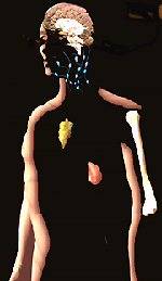
The organs of the immune system (thymus, spleen, and lymph nodes) and the organs of the neuro-immune system (adrenal gland, hypothalamus, and the cortical and subcortical brain). |
|
Interest in the limbic system remained strong through recent times, although in the last several years neuroscientists have raised questions about the looseness of some of the earlier theoretical assumptions and anatomical constructs. They are still interested in the neural substrates of emotion within the brain but have shifted their attention to the hemispheres of the cerebral cortex and to the interactions between cortical and subcortical regions. In the 1970s, neuroscientists began to concentrate on the right cortical hemisphere as the most interesting locus of emotional control.73 Roger Sperry’s award of the Nobel Prize in 1981 for his work on "cerebral laterality" (the differences between the "left" and the "right" brain and their behavioral significance) reinforced this trend, but respected neuroscientist R.W. Doty indicated in a 1989 review article that "any idea of emotion in an intact mammal being played out purely via subcortical circuitry is an unsustainable abstraction. On the other hand, the evidence is unequivocal that subcortical structures are essential for the expression of the more "primitive" emotions, and can support such expression in the absence of the neocortex."74 Current work is verifying the integrative functioning of cortical and subcortical areas (especially the amygdala) in the organism’s response to primitive emotional experiences such as fear.75 |
|
Using PET scans, scientists are in the first stages of relating different emotional states--pleasure, sorrow--to different patterns of brain activity. 

|
Functional magnetic resonance . imaging, or MRI, is another new technology that can detect the living brain at work. This is a computer-enhanced fMRI scan of a person who has been asked to look at faces. The image shows increased blood flow in the part of the visual cortex that recognizes faces. 
|
|
Powerful new imaging techniques have supported and made possible the recent emphasis on the anatomical substrates of emotion. 76 The most impressive techniques are computer assisted tomography (CAT scans), magnetic resonance imaging (MRI), positron emission tomography (PET scans), single-photon emission computed tomography (SPECT), and functional magnetic resonance imaging (fMRI). The breakthrough technology was computer assisted tomography, developed in the 1960s and 1970s, for which Allan Cormack and Godfrey Hounsfield received the Nobel Prize in 1979. The basic principle was the computer synthesis of a three-dimensional image from a series of two-dimensional "slices" taken at multiple angles (tomography) of some signal aimed at or emanating from the patient and detected outside his or her body. This principle was applied first to CAT scans where the measured property was an x-ray attenuation coefficient. The same principle was then applied to MRI imaging and PET scans, where the measured property was natural magnetization density in the first case and the concentration of an intravenously injected radioisotope in the second. 77 The newer fMRI is based on the tomographic construction of images formed by the signal differences between MRIs taken of the brain in functionally activated and non-activated states. 78 CAT scans and MRI images are now widely used in clinical settings to determine anomalies in cerebral anatomy. SPECT, PET and fMRI are valuable tools, at this point employed primarily in research settings to determine physiological and biochemical variations in brain activity, including anatomically-localized alterations in metabolism and neurochemical functioning which are visualized as they occur. 79 |
Optical
imaging camera 
The optical imaging camera allows scientists to peer even more deeply into the brain, making pictures of nerve cells working together in ensembles. A bright light shone onto the brain reflects backchanges in nerve cells activity (measured through changing colors related to water content, cell size, and amount of oxygen in the blood). These are then turned into colorful images. 
|
Computerized
photomicrographic imaging 
New technologies, like computerized photomicrographic imaging, are bringing even the microscope world of cells and genes more fully into the light of day. |
|
Many of the achievements in the neurosciences have come at the intersection of this new imaging technology with recent breakthroughs in neurochemistry.80 As one of neurochemistry’s leaders, Solomon Snyder, has said, "The glue that has brought together findings from so many different disciplines into a coherent concept of brain function is chemistry. Indeed the revolution is more precisely characterized as a revolution in ‘molecular neuroscience.’"81 Twenty years ago, Snyder was among those neurochemists who succeeded in identifying opium-like molecules in the brain (variously called "enkephalins," "endorphins," or sometimes just "endogenous opioids") that helped regulate the sensation of pain. Endogenous opioids are a type of "neurotransmitter," a long-studied class of biochemical substances that convey messages from nerve fiber endings to other biological receptors, whether nerve, muscle or gland. Neurochemists were able to identify specific opiate "receptor sites" where the endogenous opioids normally attach but at which they are sometimes displaced by exogenous competitors such as morphine. Using photographic techniques that take pictures of samples incorporating radioactive materials and high power microscopy, scientists found large concentrations of these receptor sites in areas of the brain (in the limbic system) specifically associated with pain perception and other forms of emotional regulation.82 More recently and with the help of PET scan and fMRI technology, neuroscientists have been able to confirm the dense distribution of opiate receptors in the structures of the limbic system and especially in the amygdala. Neuroscientists thus seem to be closing in on both the biochemical mechanisms and the anatomical architecture of emotional expression in specific structures of the brain. |

 |
Neurotransmitters Different parts of your brain share information and organize plans for action through a code system that involves both chemistry and electricity. Chemicals called neurotransmitters" are emptied from tiny sacs into the space between nerve cells. These chemicals cross that space and bind to receptors on other nerve cells. The binding process triggers an electrical stimulus in the receiving cells, that starts the whole process of chemical release all over again. | |
|
In perhaps the most exciting development of all, a new field has emerged which is starting to combine the latest in the neurosciences with the latest in immunology to provide the scientific basis for understanding relationships between emotions and disease once explored only in clinical settings. Not yet possessing a generally agreed upon name, this new field has been able to demonstrate previously unsuspected but now verifiably direct connections between the immune system and the neuroendocrine system. The field developed in two waves. The first wave, rising in the late seventies and early eighties, was generally called "psychoneuroimmunology" (PNI). Its roots could in some sense be traced back to the pioneering studies of the Russian immunologist S. Metal’nikov at the Pasteur Institute in Paris in the 1920s and 1930s and to the considerable work in the Soviet Union from the 1920s through the 1950s on psychologically conditioned immunobiological effects. The field really began to take shape around 1980 under the combined leadership of the Americans George Solomon, Novera Herbert Spector and Robert Ader, the Swiss Hugo Besedovsky, and the Russian Elena A. Korneva. 83 Although each of these leaders came from a different discipline and contributed different specific expertise (Ader, for example, was an experimental psychologist, Solomon was a psychiatrist and Besedovsky was an endocrinologist), they all agreed on the need to break down the barriers that until then had artificially separated immunology as a field from endocrinology and the neurosciences. As Ader and his colleagues put the point in 1987, "In our view, the attempt to understand immunity as an adaptive process that is independent of and can be studied in isolation from other integrated adaptive processes is, in its extreme form, a restrictive and restricting paradigm." 84 |
Activation
of T-cells 
When a foreign toxin or bacteria, called an antigen, enters the body, immune system cells race to the site of invasion. These cells are called lymphocytes (T and B cells) and macrophages. Receptors on the surface of these cells recognize and bind to the invader. The binding process triggers the production of chemical signals called interleukins. Interleukins allow immune cells to mature, communicate with each other, and to make antibodies and other substances that remove the invader. |
At the same time that interleukins (sometimes called lymphokines and monokines) allow immune cells to signal one another, they also allow immune cells to signal the brain--and vice versa. | |

|
Beginning in the late 1980s, the second wave was marked by the recruitment of molecular neuroscientists. This phase does not yet have a fixed name, although "neuroimmunomodulation" (NIM) is widely accepted, while some leaders prefer simply "neuro-immune interactions." Some of the scientists recruited to the field during this phase were wary of PNI and remained skeptical until they were persuaded by "harder" evidence that the immune and neuroendocrine systems are in fact in close and bi-directional communication and, indeed, "talk" to each other all the time. A short list of discoveries early in the second wave includes the following: demonstration of direct microanatomical contacts between the nervous and the immune systems discovery that anatomical lesions in or the electrical stimulation of parts of the brain influence antibody production in the spleen and lymph nodes; identification of receptor sites for neuroendocrine hormones and neurotransmitters on cells of the immune system. The "clincher" was the repeated proof in several different animal models that interruptions of these communications on a genetic, surgical, or pharmacological basis, lead to increased susceptibility to inflammatory diseases like arthritis. The converse is now also being shown, that too much responsiveness of these systems leads to enhanced susceptibility to infection. Now it is certain that particular molecules of the immune system (cytokines or interleukins) signal areas of the brain directly as well as exert influences on peripheral parts of the nervous system such as the vagus nerve. This rigorously demonstrated "cross-talk" between the immune and neuroendocrine systems has won over neuroscientists and gained converts among the immunologists themselves. Even more important, it provides the scientific basis for understanding--at long last--how emotions can in fact influence the onset, course, and remission of disease. |
|
Two very different signs of enthusiasm and "arrival" already mark the 1990s: the inclusion of an entry on "Neuroendocrine Regulation of Immunity" in the 1992 Encyclopedia of Immunology 85 and the featuring of psychoneuroimmunolgy as a central theme in Bill Moyers's 1993 best seller, Healing and the Mind. 86 The first indicated the acceptance of the new field within the mainstream of previously resistant immunology and the second demonstrated popular fascination with the emerging inter-discipline. Moyers and many of his readers seized upon the new field as seeming to validate long-suspected but frequently denied connections between emotions and disease. A spate of high-level international scientific conferences marked by unusual energy and bold proclamations have added to the sense of excitement. The proceedings of one of these was published in 1994 as volume three of the "Hans Selye Symposia on Neuroendocrinology and Stress." 87 The editors of the Selye volume capture the current mood: |
|
The interaction of the nervous, endocrine and immune systems is only now being considered seriously. This field represents a novel, multidisciplinary approach in Biological Sciences. Even the name of the field has not been settled as yet and there are debates going on with regards to the proper term. . . . Modern science is equipped with powerful research tools which make it feasible to advance quickly in this complex multidisciplinary field, with the aim of understanding the whole organism, rather than trying to analyze restricted areas. The developments are spectacular, indeed, and the new insights gained . . . have already advanced our understanding of certain human diseases, such as autoimmune disease, inflammatory diseases, nervous and endocrine abnormalities and the influence of behavioral factors and of aging on the immune response and disease. We sincerely hope this volume will contribute to the understanding and acceptance of this brave new area of scientific enquiry. |
|
It may be that this "brave new area" will finally validate long held beliefs about emotions and disease that we in the West have been grappling with for at least two millennia. |

|